Pelvic Anatomy Ligaments / Female Pelvis Diagram Anatomy Function Of Bones Muscles Ligaments : The ilium, ischium and the pubic bone.. The pelvis's frame is made up of the bones of the pelvis, which connect the axial skeleton to the femurs, and therefore acts in weight bearing of the upper body. • posterolateral wall—piriformis and coccygeus muscles. Inherent stability of the pelvis is provided by ligaments. This image shows the posterior back view of the female pelvic brim (the bones and ligaments that forms the pelvic region in the female) showing: They form what can be described as a basket weave formation, in order to create strength and tensegrity within the structure.
The pelvis is held together by three principal ligaments: The pelvic ligaments are strong, thick bands of fibrous tissue that connect the pelvic bones. It is not considered a true ligament in that it does not. The femoral ligaments act to stabilize the ball and socket joint of the hip, connecting to the ilium and the ischium. Iliolumbar, sacrotuberous and sacrospinous ligaments.
Bones Ligaments Joints Atlas Of Anatomy from doctorlib.info The pectineal ligament, sometimes known as the inguinal ligament of cooper, is an extension of the lacunar ligament. Bones and ligaments of the female pelvis. The sacral ligaments are responsible for the major connection between the three bones of the pelvis. The suspensory ligament of the ovary, also infundibulopelvic ligament (commonly abbreviated ip ligament or simply ip), is a fold of peritoneum that extends out from the ovary to the wall of the pelvis. They form what can be described as a basket weave formation, in order to create strength and tensegrity within the structure. The pelvic girdle, also known as the hip bone, is composed of three fused bones: The enclosed space between the inlet and outlet is called the true pelvis, with. Pelvic bone and ligaments anatomy.
Iliolumbar, sacrotuberous and sacrospinous ligaments.
Inherent stability of the pelvis is provided by ligaments. Lets get deeper into the musculoskeletal anatomy of the hip and look at the bones and bony bits of the pelvis, and the ligaments that attach here and hold it. • anterolateral wall—hip bone and obturator internus muscles. We hope you can get the exact. The cardinal ligament (cl) still requires more precise anatomical mapping. The pelvic girdle, also known as the hip bone, is composed of three fused bones: This image shows the boundaries of the pelvic area formed of the pelvic bones and ligaments showing: The cardinal ligament is a paired thickening of the parametrium and pelvic fascia at the base of the broad ligament, which extends between the cervix and vaginal fornix medially to the sidewall of pelvis laterally. Pelvic bone and ligaments anatomy. These ligaments are categorized into four groups: It runs on the pectineal line of the pubic bone. The broad ligament is a flat sheet of peritoneum, associated with the uterus, fallopian tubes and ovaries. The outlet is formed by the pubic arch, ischial spines, sacrotuberous ligaments, and the coccyx.
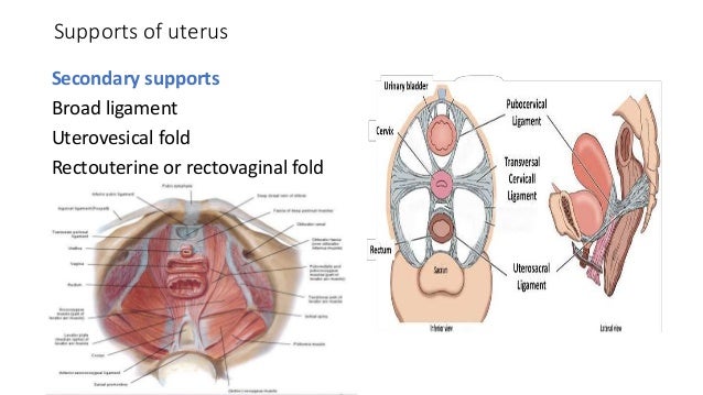
• posterior boundary—sacrum and coccyx. These ligaments are categorized into four groups: • muscles and ligaments form a pelvic floor. • located inferior to the pelvic brim. Broad ligament the broad ligament supports the uterus, fallopian tubes, and ovaries.
Pelvic Floor Disorders Pelvic Girdle Pain And Symphysis Pubis Dysfunction Following Sports Injury Caring Medical Florida from www.caringmedical.com We aim to elucidate the anatomy of the cl and the roles it plays in gynecological surgery. Some sources consider it a part of the broad ligament of uterus while other sources just consider it a termination of the ligament. This image shows the boundaries of the pelvic area formed of the pelvic bones and ligaments showing: The pelvic girdle, also known as the hip bone, is composed of three fused bones: • posterior boundary—sacrum and coccyx. Inherent stability of the pelvis is provided by ligaments. Über 7 millionen englischsprachige bücher. The pelvic girdle and pelvic spine.
The pelvis is held together by three principal ligaments:
This will be explored further on. Pelvic bone and ligaments anatomy. We aim to elucidate the anatomy of the cl and the roles it plays in gynecological surgery. This image shows the posterior back view of the female pelvic brim (the bones and ligaments that forms the pelvic region in the female) showing: We think this is the most useful anatomy picture that you need. Anterior superior iliac spine 6. • also known as pelvic cavity. Bones and ligaments of the female pelvis. Ligaments connect one bone to another and provide important stability. The pelvis is a boney structure at the base of the lumbar spine. The broad ligament can be further divided into three components. This image shows the boundaries of the pelvic area formed of the pelvic bones and ligaments showing: It extends to both sides of the pelvic wall.
The named ligaments of the pelvis mostly arise from the sacrum and attach to varying segments of the pelvic bone. Joints and ligaments of the pelvis the two sacroiliac joints are synovial joints, and are further strengthened by the very strong posterior sacroiliac ligaments which run along the posterior aspect of the joint. Anatomynote.com found pelvis and ligaments front view from above male from plenty of anatomical pictures on the internet. Über 7 millionen englischsprachige bücher. The femoral ligaments act to stabilize the ball and socket joint of the hip, connecting to the ilium and the ischium.
Surgical Anatomy Of Pelvis from image.slidesharecdn.com The outlet is formed by the pubic arch, ischial spines, sacrotuberous ligaments, and the coccyx. It is usually divided into two separate anatomic regions: The inlet to the pelvic canal is at the level of the sacral promontory and superior aspect of the pubic bones.; It is not considered a true ligament in that it does not. The ligaments that pass between the sacrum and the ischium, which is the lower rear part of the pelvis; The pelvis is held together by three principal ligaments: These ligaments are important stabilizers. Iliolumbar, sacrotuberous and sacrospinous ligaments.
These ligaments are important stabilizers.
Broad ligament the broad ligament supports the uterus, fallopian tubes, and ovaries. The enclosed space between the inlet and outlet is called the true pelvis, with. The suspensory ligament of the ovary, also infundibulopelvic ligament (commonly abbreviated ip ligament or simply ip), is a fold of peritoneum that extends out from the ovary to the wall of the pelvis. Cardinal ligament and the uterosacral ligaments provide apical support for the uterus and upper vagina. They form what can be described as a basket weave formation, in order to create strength and tensegrity within the structure. Other ligaments attached to bony pelvis include the sacrococcygeal ligaments, pubic symphysis ligaments, and endopelvic fascia ligament. Ligaments connect one bone to another and provide important stability. This is part of the forced closure method that the pelvis adopts in order to keep itself secure. It is usually divided into two separate anatomic regions: The pelvis itself is a paired composite structure made up by three bones (ilium, ischium and pubis) that articulates with the sacral part of the axial spine. Those that connect the ilium to the sacrum; Some sources consider it a part of the broad ligament of uterus while other sources just consider it a termination of the ligament. Über 7 millionen englischsprachige bücher.


0 Komentar